Home
/ Back Bones Diagram - Human Bones Anatomy Anatomy Bones Human Bones : It is particularly interesting for physiotherapists.
Back Bones Diagram - Human Bones Anatomy Anatomy Bones Human Bones : It is particularly interesting for physiotherapists.
Back Bones Diagram - Human Bones Anatomy Anatomy Bones Human Bones : It is particularly interesting for physiotherapists.. Bones of the pelvis and lower back. In the back and elsewhere in the body, tendons attach muscles to bones. 12 photos of the human back bone chart. The human back extends from the buttocks to the posterior portion of the neck and shoulders. Fishbone diagrams, aka ishikawa diagrams are used across various industries to analyze causes and their effect.
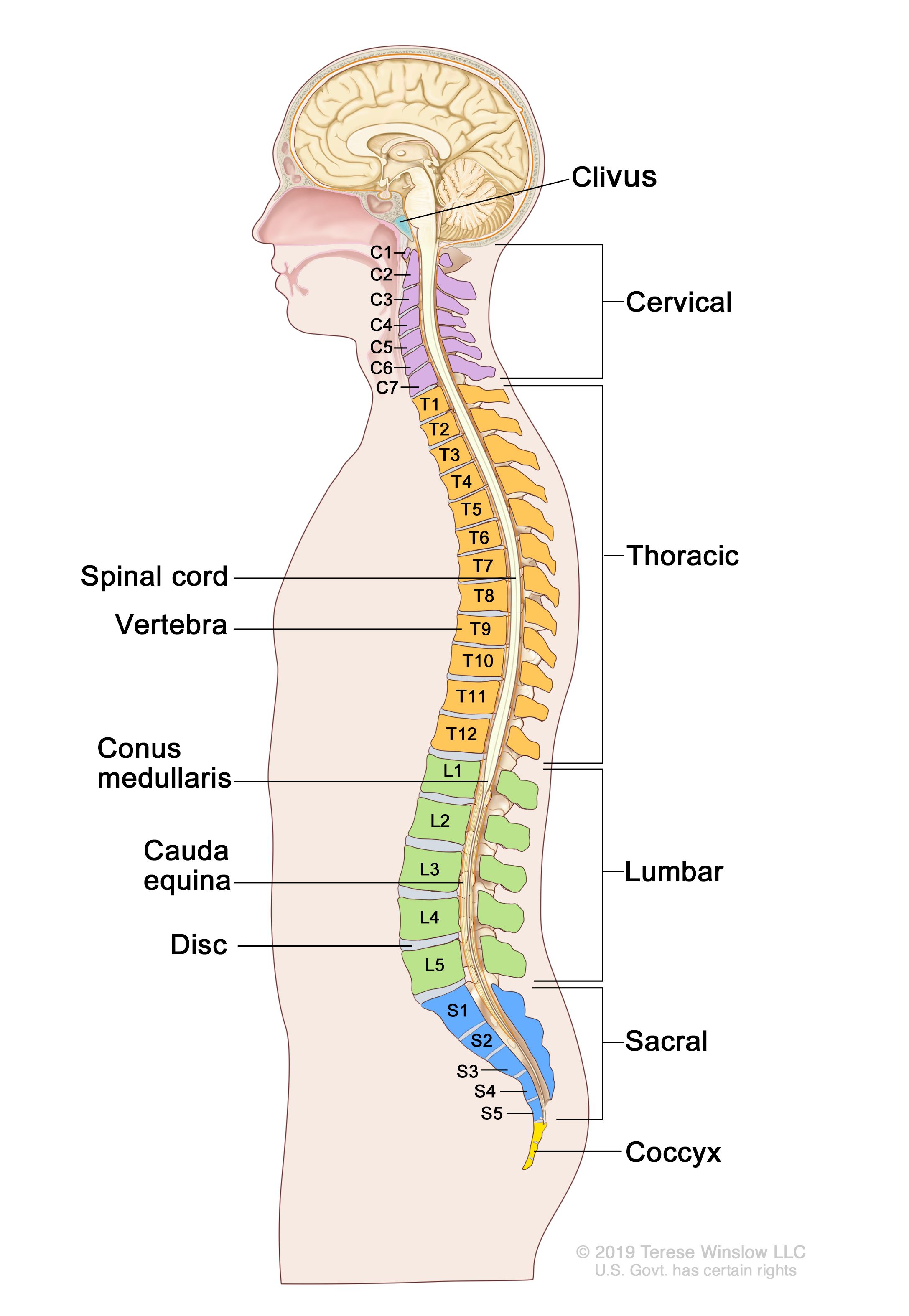
So many patients receive diagnostic imaging reports full of terms and anatomical locations which are unknown and mysterious to them. Spinal anatomy and back pain. Your lower back contains 5 vertebral bones stacked above each other with intervertebral discs in between. Exercises can strengthen the core muscles that support the spine and. Its appearance is different from the other spinal vertebrae.
Anatomy Of The Back Spine And Back Muscles Kenhub from thumbor.kenhub.com Related posts of human back bones diagram bone structure birds. Bones, discs, and joints in your lower back. The disks that cushion vertebrae may compress with age or injury, leading to a herniated disk. Spinal anatomy and back pain. At the same time the bones grow larger by growing back into the growth plates. The notochord present in the embryonic stage is replaced by the vertebral column. The spine anatomy is a complex structure. Human backbone diagram, bone, human backbone diagram.
Seven cervical vertebrae in the neck, twelve thoracic vertebrae in the torso and five lumbar vertebrae in the lower back.
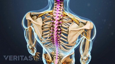
Skeletal diagrams can also be used to show bone development or growth which begins en utero. Bones, discs, and joints in your lower back. Nerves of the abdomen lower back and pelvis. It contains the osteology, arthrology and myology of the spine and back. Exercises can strengthen the core muscles that support the spine and. The disks that cushion vertebrae may compress with age or injury, leading to a herniated disk. Spinal vertebrae bone spine vertebra toracica spinal cord spine structure back diagram spine sections spinal cord vertebrae spinal structure health diagram. A posterior view skeletal diagram provides a back view of the human skeleton. Key parts of your spine include vertebrae (bones), disks, nerves and the spinal cord. The column can be divided into five different regions, with each region characterised by a different vertebral structure. This process continues until the end of puberty, when the growth plate stops growing and the bones fuse permanently into a single bone. Each typical vertebra consists of a body, an arch and three processes that stem from. The first seven bones (vertebrae) of your spine form your neck.
Anatomical diagrams of the spine and back. It is also known as the vertebral column. Can you feel the bumps of your vertebrae along your back? Nerves of the abdomen lower back and pelvis. We think this is the most useful anatomy picture that you need.
Definition Of Backbone Nci Dictionary Of Cancer Terms National Cancer Institute from nci-media.cancer.gov The bones of the pelvis and lower back work together to support the body's weight, anchor the abdominal and hip muscles, and protect the delicate vital organs of the vertebral and abdominopelvic cavities. The spine anatomy is a complex structure. The first seven bones (vertebrae) of your spine form your neck. At the same time the bones grow larger by growing back into the growth plates. The lumbar spine is the lower back that begins below the last thoracic vertebra (t12) and ends at the top of the sacral spine, or sacrum (s1). The column can be divided into five different regions, with each region characterised by a different vertebral structure. The vertebral column is a part of the axial skeleton, which comprises the skull, ribs and sternum other than the vertebral column. They help support particular bones and make them move.
Urinary system of the lower torso.
It also covers some common conditions and injuries that can affect the back. At the back of each bone in the spine (vertebra) are bony points called processes, which muscles attach to. A posterior view skeletal diagram provides a back view of the human skeleton. Vertebrae are the structural constituents of the spine.there are 33 vertebrae in total; These bones are connected at the back with specialized joints. It is designed to be incredibly strong, protecting the highly sensitive nerve roots, yet highly flexible, providing for mobility on many different planes. They help support particular bones and make them move. Skeletal diagrams can also be used to show bone development or growth which begins en utero. Anatomynote.com found anatomy of back muscles diagram from plenty of anatomical pictures on the internet. Lateral labeled diagram of the human vertebral spinal column showing vertebrae numbering order and the 5 different regions of the spine. The column can be divided into five different regions, with each region characterised by a different vertebral structure. The bones of the pelvis and lower back work together to support the body's weight, anchor the abdominal and hip muscles, and protect the delicate vital organs of the vertebral and abdominopelvic cavities. It is opposite from the chest, and the vertebral column runs down the back.
The spine or backbone consists of 26 small bones or vertebrae. Vertebrae are the structural constituents of the spine.there are 33 vertebrae in total; This process continues until the end of puberty, when the growth plate stops growing and the bones fuse permanently into a single bone. Key parts of your spine include vertebrae (bones), disks, nerves and the spinal cord. Its appearance is different from the other spinal vertebrae.
Spinal Anatomy And Back Pain from embed.widencdn.net The lumbar spine connects to the thoracic spine above and the hips below. The vertebrae, which stack like spools of thread, support the back and protect the spinal cord. Your lower back contains 5 vertebral bones stacked above each other with intervertebral discs in between. Daniel nelson on january 1, 2019 2 comments 🔥! We think this is the most useful anatomy picture that you need. The human back extends from the buttocks to the posterior portion of the neck and shoulders. The first seven bones (vertebrae) of your spine form your neck. For more anatomy content please follow us and visit our website:
A tough, springy disc of cartilage sits between the vertebrae of your spine.
It is designed to be incredibly strong, protecting the highly sensitive nerve roots, yet highly flexible, providing for mobility on many different planes. Skeletal diagrams can also be used to show bone development or growth which begins en utero. Spinal anatomy and back pain. The atlas is a ring of bone made up of two lateral masses joined at. Anatomical diagrams of the spine and back. The vertebral column is a series of approximately 33 bones called vertebrae, which are separated by intervertebral discs. Its appearance is different from the other spinal vertebrae. A lateral view skeletal diagram offers a side view of the human skeleton. Fishbone diagrams, aka ishikawa diagrams are used across various industries to analyze causes and their effect. The vertebral column is a part of the axial skeleton, which comprises the skull, ribs and sternum other than the vertebral column. At the back of each bone in the spine (vertebra) are bony points called processes, which muscles attach to. The basics of back pain and spinal anatomy. Vertebrae are the structural constituents of the spine.there are 33 vertebrae in total;
:background_color(FFFFFF):format(jpeg)/images/library/12063/primary-authochtone-back-muscles_english.jpg&description=Back Bones Diagram - Human Bones Anatomy Anatomy Bones Human Bones : It is particularly interesting for physiotherapists.)